
http://www.whitman.edu/biology/vpd/main.html This is a website of a fetal pig dissection
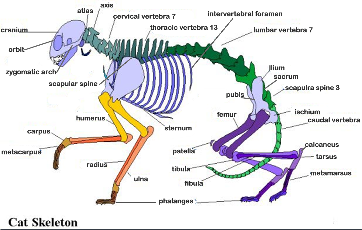
click here to Print Cat Skeleton with Lables ** set page to print landscape
Click Here to Print Cat Skeleton without lables ** set page to print landscape
![]()
Some of my notes from reading the lab manual
Vestibule- area between lips and gums
Hard palate- roof of mouth
Soft palate
Pharynx
Epiglottis- region on the lower part of tongue – closes when animal swallows
Glottis- the hole in the epiglottis- opening to the larynx
Esophagus
Parotid gland- salivary gland
Thymus gland- controls metabolism growth and development hormones
Thyroid gland covers trachea (2 lobes) development and maintenance of immune system
Trachea- has cartilaginous support rings
Heart- is in pericardial sac
Lungs-
Diaphragm
Liver- four lopes secretes bile maintaining composition of blood
Stomach- bean shaped organ
Pancreas between stomach and small intestine regulates insulin
Spleen- below stomach
Umbilical vein – criteria
Spiral intestine
Small intestine – 3 regions, duodenum- along the pancreases, jejunum and ileum
Villi- small projections from wall of intestine
Large intestine (colon) ileum
Bronchi- tube of the lungs divide into smaller branches
Vocal cords – inside the larynx
Pleural cavities- contains lungs and pericardial sac
Blood vessels- transports: water oxygen CO2 metabolic wastes, and hormones
Pulmonary circuit- blood flow to and from heart and lungs
Systemic circuit- blood to the rest of the body
4 heard chambers
Right atria
Left atria
Right ventricle
Left ventricle
Coronary vessels- separates ventricles of the heart- supplies hear with blood to function
Vena cava- large blue vein at the top (superior) and bottom (interior) of the heart
Pulmonary trunk- between left and right atria
Right & Left pulmonary arteries branch from pulmonary trunk
Right and left pulmonary veins return from the lungs to the heart with oxygenated blood
Ductus arterious- a large but short vessel connecting the pulmonary trunk and the aorta- for fetal blood oxygenation
Branchiocephalic artery- branches from aortic arch
Right subclavian artier going into the right fore limb
Carotid trunk branch through the neck and head
Left subclavian artery- branches through the left shoulder
Clavian vein becomes the brachiocephalic vein that junctures to the superior vena cava
Left and right common carotid arteries- what brachiocephalic artery splits into immediately
Internal and external jugular veins- on either sided of the neck
Superior mesenteric artery below the celiac artery and branches to the pancreas and duodenum of the small intestine
Renal arteries go to the kidneys
The external iliac arteries branch to each hind limb and leg
Common iliac vein drains back from the legs
I am glad to everyone who has visited my website this semester. I hope my notes have helped you.